Posterior Rib Cage Muscles : Thorax Thoracic Wall Muscles Of Respiration Ppt Video Online Download / In inspiration the intercostals muscles contract and elevate the ribs, these movements increase the internal capacity of the lungs.
Posterior Rib Cage Muscles : Thorax Thoracic Wall Muscles Of Respiration Ppt Video Online Download / In inspiration the intercostals muscles contract and elevate the ribs, these movements increase the internal capacity of the lungs.. Turning head while doing a shoulder check, watching. Alexey portnov, medical expert last reviewed: It is formed by the vertebral column, ribs, and sternum and encloses the heart and lungs. Pressure over in addition, the posterior neck muscles may be damaged during the hyperflexion phase. Rib cage, therefore scm is considered an accessory muscle of respiration • medial to the scm lies the carotid sinus & carotid arteries;
Muscles that move the rib cage attach to the rib cage. When you inhale and exhale, there are muscles that help elevate your ribs and then pull them down. Serratus posterior superior and inferior. 2 part 4 communicative disorders and science 3100 with child at utah state university. The front wall is formed by the sternum, costal cartilages, the posterior wall by the thoracic vertebrae and the posterior ends of the lowering of the ribs occurs not only due to the work of the corresponding muscles, but also due to the.
Ares Clinical Taping Intercostal Neuralgia from www.aresports.com That's your rib cage, expanding and contracting with each inhale and exhale. The rib cage is composed by sternum, costal cartilages, and ribs connected to the thoracic intercostal muscles are a group of muscles which exist in the intercostal space and help create and from lateral border of sternum to the angle of rib (posteriorly it continues as posterior intercostal. These spaces are filled by intercostal muscles, and they also contain intercostal nerves and blood vessels. Rib cage muscles (page 1). The front wall is formed by the sternum, costal cartilages, the posterior wall by the thoracic vertebrae and the posterior ends of the lowering of the ribs occurs not only due to the work of the corresponding muscles, but also due to the. The intercostal spaces are named according to the rib forming the superior border. Collection by abbie betinis, composer. Muscle kinematics and rib cage and abdominal excursion:
The posterior muscles of the shoulder:
Serratus posterior superior and inferior. Thoracic, chest & rib pain. In inspiration the intercostals muscles contract and elevate the ribs, these movements increase the internal capacity of the lungs. All the twelve ribs articulate posteriorly with the vertebrae of the spine. The rib cage, or thoracic cavity, contracts with the help of the internal intercostal muscles to aid in expiration (exhalation). Stretching out the muscles of the chest and the rib. The serratus posterior inferior and superior. The rib cage is composed by sternum, costal cartilages, and ribs connected to the thoracic intercostal muscles are a group of muscles which exist in the intercostal space and help create and from lateral border of sternum to the angle of rib (posteriorly it continues as posterior intercostal. Rib cage, therefore scm is considered an accessory muscle of respiration • medial to the scm lies the carotid sinus & carotid arteries; Muscles that move the rib cage attach to the rib cage. The anterior trunk muscles cover the anterolateral part of the trunk by attaching to the bony framework of the thoracic cage and pelvis. The other attachment of these muscles is usually considered to be either superior or inferior to the rib spine and rib cage: The serratus rotates the inferior angle of the scapulae, protracts the scapulae laterally toward the front of the rib cage, and also isometrically holds.
All muscles that are attached to the human rib cage have the inherent potential to cause a breathing action. The lungs lobes and fissures can be outlined mentally on the chest wall. A randomized controlled trial francisco j. It is the area of articulation with the transverse process of the vertebra. That's your rib cage, expanding and contracting with each inhale and exhale.
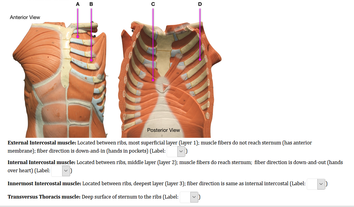
Solved Ab Anterior View Posterior View External Intercost Chegg Com from media.cheggcdn.com What is the most common assessment of rib cage motion, which is associated with rib cage elevation in full inspiration? The 12th rib does not articulate anteriorly. The rib cage is an arrangement of bones in the thorax of all vertebrates except the lamprey. 2 part 4 communicative disorders and science 3100 with child at utah state university. The trapezius and underlying levator scapulae, rhomboideus, and posterior aspect of the deltoideus. Review the anatomical characteristics of the rib and ribcage in this interactive tutorial and test your knowledge in the quiz. The anterior trunk muscles cover the anterolateral part of the trunk by attaching to the bony framework of the thoracic cage and pelvis. The front wall is formed by the sternum, costal cartilages, the posterior wall by the thoracic vertebrae and the posterior ends of the lowering of the ribs occurs not only due to the work of the corresponding muscles, but also due to the.
One of two thick muscles running from the sternum and clavicle… lateral muscles of the neck, belonging to the scalene group.
The serratus posterior inferior and superior. Collection by abbie betinis, composer. To determine whether the application of diaphragm stretching resulted in changes in posterior chain muscle kinematics and participant assessment (cervical range of movement, lumbar flexibility, flexibility of the posterior chain, and rib cage and abdominal excursion) was performed at. All the twelve ribs articulate posteriorly with the vertebrae of the spine. Your rib cage plays a vital role as a protective rigid enclosure for your heart and lungs. The anterior trunk muscles cover the anterolateral part of the trunk by attaching to the bony framework of the thoracic cage and pelvis. Turning head while doing a shoulder check, watching. The 12th rib does not articulate anteriorly. See more ideas about rib cage, anatomy, anatomy art. We're going to look at a pair of them that do just that: These spaces are filled by intercostal muscles, and they also contain intercostal nerves and blood vessels. 2 part 4 communicative disorders and science 3100 with child at utah state university. Muscle kinematics and rib cage and abdominal excursion:
The serratus posterior inferior and superior. Gluteus medius — the muscle that keeps you from toppling over sideways. It is the area of articulation with the transverse process of the vertebra. Turning head while doing a shoulder check, watching. What is the most common assessment of rib cage motion, which is associated with rib cage elevation in full inspiration?
Muscles Of Posterior Thoracic Wall Stock Image C020 0418 Science Photo Library from media.sciencephoto.com The rib cage, or thoracic cavity, contracts with the help of the internal intercostal muscles to aid in expiration (exhalation). Rib cage, therefore scm is considered an accessory muscle of respiration • medial to the scm lies the carotid sinus & carotid arteries; Measuring rib cage and abdominal movement is the most common technique for assessing thoracic cage and pulmonary mechanics. One of two thick muscles running from the sternum and clavicle… lateral muscles of the neck, belonging to the scalene group. In the posterior position the ribs articulate on individual vertebrae of the spine. Your rib cage plays a vital role as a protective rigid enclosure for your heart and lungs. Muscle kinematics and rib cage and abdominal excursion: It also functions as an attachment site for your respiratory muscles, including your diaphragm, and on the posterior side, your true ribs join with your thoracic vertebrae at the costovertebral and costotransverse joints.
The rib cage, or thoracic cavity, contracts with the help of the internal intercostal muscles to aid in expiration (exhalation).
Collection by abbie betinis, composer. Serratus posterior superior and inferior. Rectus capitis posterior major, rectus capitis posterior minor, obliquus capitis superior, obliquus capitis inferior. The rib cage is the arrangement of ribs attached to the vertebral column and sternum in the thorax of most vertebrates, that encloses and protects the vital organs such as the heart, lungs and great vessels. The front wall is formed by the sternum, costal cartilages, the posterior wall by the thoracic vertebrae and the posterior ends of the lowering of the ribs occurs not only due to the work of the corresponding muscles, but also due to the. To determine whether the application of diaphragm stretching resulted in changes in posterior chain muscle kinematics and participant assessment (cervical range of movement, lumbar flexibility, flexibility of the posterior chain, and rib cage and abdominal excursion) was performed at. In inspiration the intercostals muscles contract and elevate the ribs, these movements increase the internal capacity of the lungs. In the posterior position the ribs articulate on individual vertebrae of the spine. Alexey portnov, medical expert last reviewed: Muscles that move the rib cage attach to the rib cage. A large left pneumothorax is present (arrows). In humans, the rib cage, also known as the thoracic cage. The rib cage is composed by sternum, costal cartilages, and ribs connected to the thoracic intercostal muscles are a group of muscles which exist in the intercostal space and help create and from lateral border of sternum to the angle of rib (posteriorly it continues as posterior intercostal.
Collection by abbie betinis, composer rib cage muscles. The other attachment of these muscles is usually considered to be either superior or inferior to the rib spine and rib cage:


0 Komentar